| Muscle Modeler | |||
| Objective | quantification of architectural parameters (e.g., fascicle length, pennation angle, physiological cross-sectional area and volume) and 3D reconstruction of surface and volume geometry | ||
| Methods | dissection and digitization of cadaveric specimens (MicroScribe G2X) and ultrasonography | ||
| Outputs | architectural analysis and geometric mesh (triangular and tetrahedral) | ||
| Applications | cadaveric studies : extensor/flexor forearm muscles, supraspinatus/infraspinatus, pectoralis major, vastusmedialis, piriformis, sacral nerves, ACL/PCL ligaments, humerus tendon rupture. in-vivo studies: supraspinatus/infraspinatus (current) | ||
| Development | C/C++, MFC, OpenGL, BLAS, LAPACK, TAUCS, MS Windows platforms | ||
| Keywords | dissection, digitization, cubic spline, voronoi-tessellation, level-set method, tetrahedralization, Laplacian surface, linear/non-linear least squares, finite element method | ||
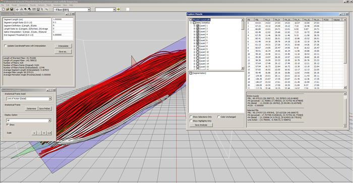
| Screenshot examples | ||
| Mainframe and examples of dialog-based control (editing digitized points, anatomical frame estimation and fascicular investigation) | ||
| Main features include basic geometric manipulation, architectural analysis, surface/volume extraction, FEM-based muscle deformation, geometric deformation, virtual ultrasound imaging, digitization error reduction, geometric cross-matching of architectures, 3D registration and etc. | ||
|
|
||
|
|
||
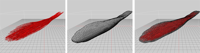
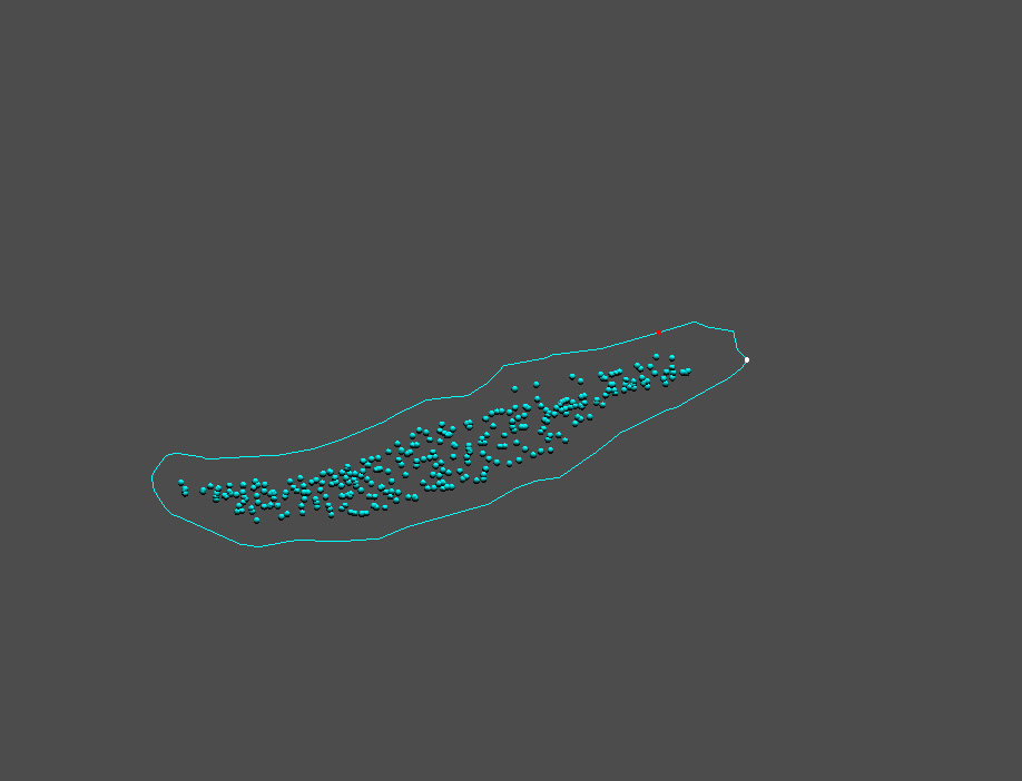
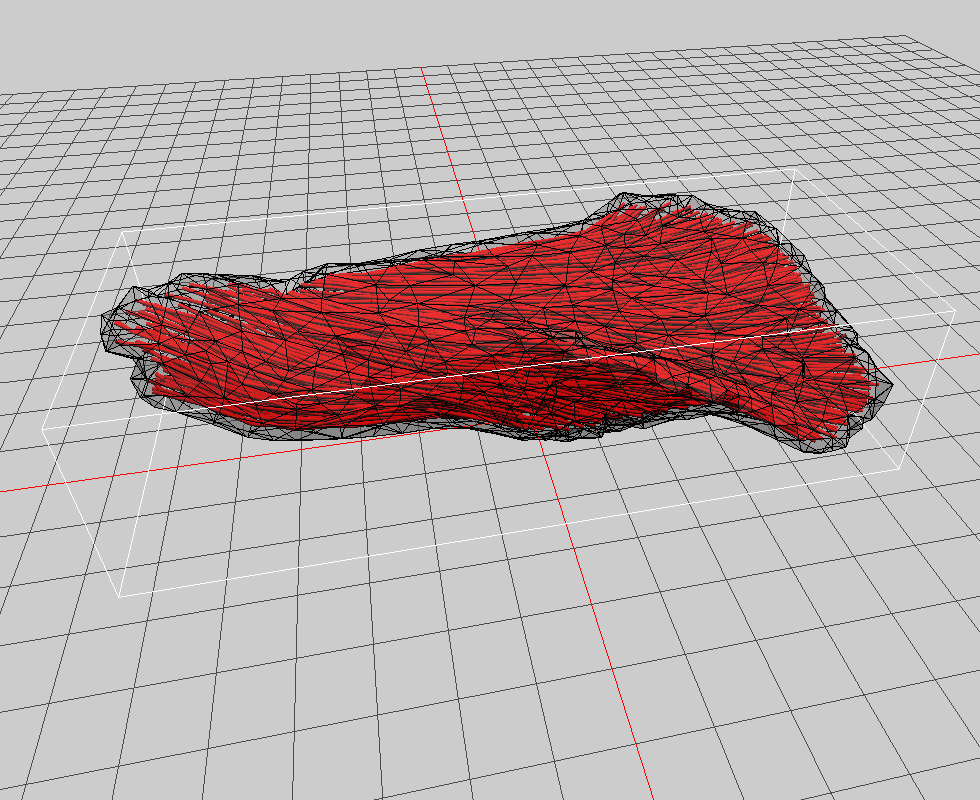
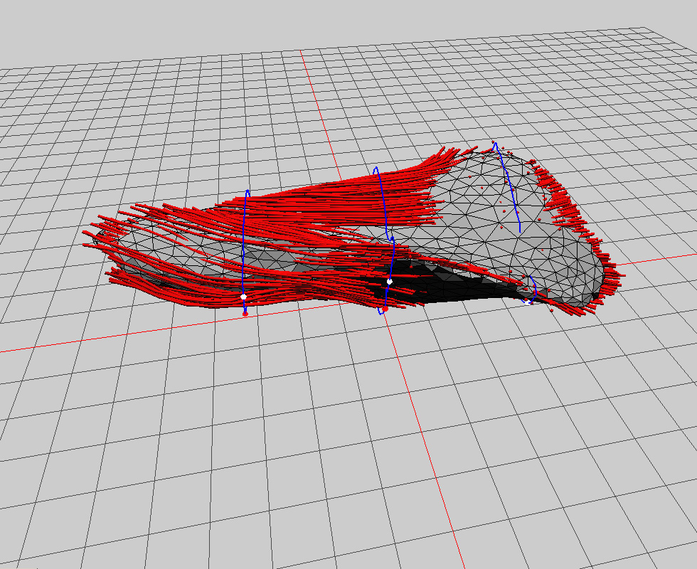
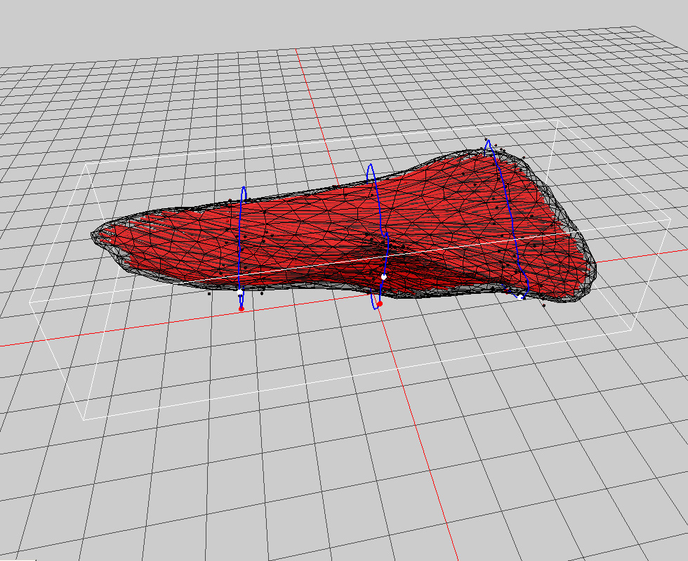
| Reconstruction of surface and volume geometry | |||
| Digitized fascicles are wrapped around by the closed surface that is tracked using the level-set method. Associated implicit functions are determined based on the estimated anatomical cross-sectional area of each fascicle (i.e., elliptical and piecewise cylinders). Those functions are carefully blended by using the locally-weighted interpolation, which controls smoothness and prevents undesired geometric defects (e.g., hole, gaps, concavity). Brachioradialis is shown in figures. | |||
|
|||
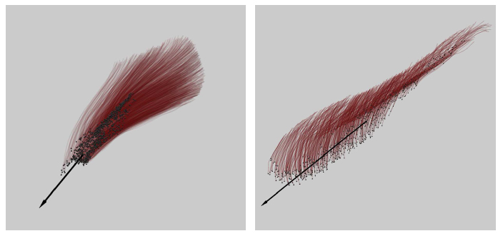
| Estimation of line of action | ||
| Linear regression is used to determine line of action (intra-and extra muscular tendon direction) of pennate muscles. (a. supraspinatus and b. vastusmedialis) | ||
 |
||
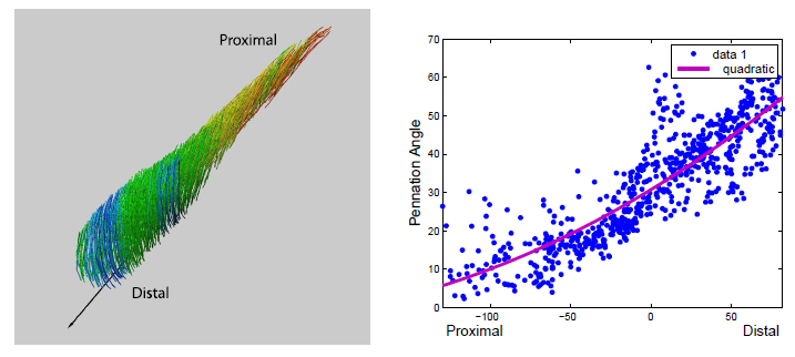
| Assessment of intramuscular variation of pennation angle | ||
| It is observed that muscles may have strong pattern of pennation angle distribution with respect to anatomical axis. Proximodistal, lateromedial and superficial to deep axes are accounted for presented studies. | ||
 |
||
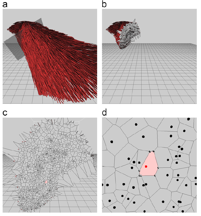
| Estimation of physiological cross-sectional area | ||
| Voronoi-tessellation is used to approximate cross-sectional area of each fascicle. This method iteratively computes all fascicles and their points. Our study uses 30-60K re-sampled points, depending on architectural complexity or size. | ||
 |
||
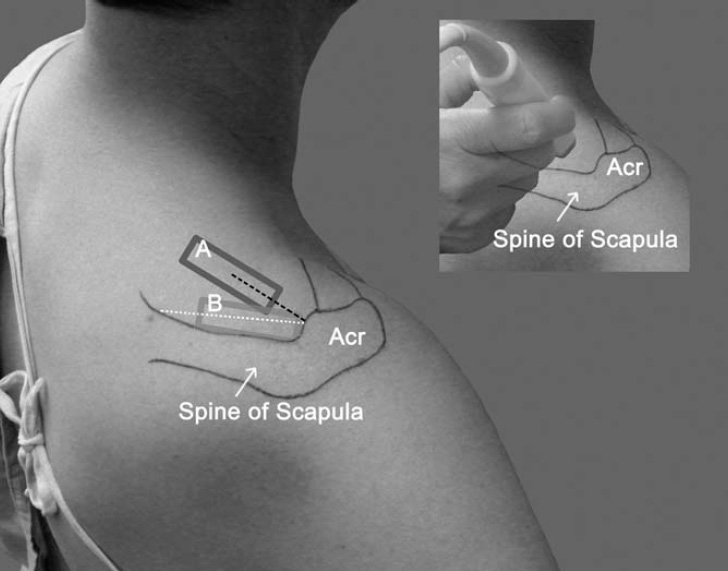
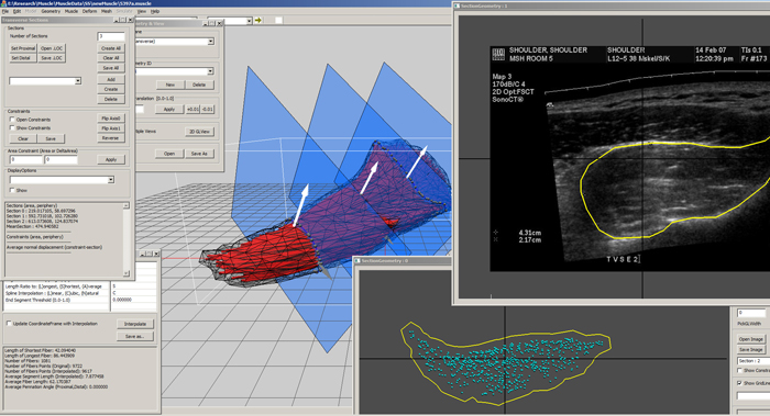
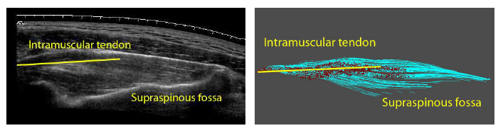
| Integration of ultrasonographic assessment for in-vivo architectural modeling | ||
| Our study stresses that anatomical details are needed to enhance reliability and reproducibility of ultrasonography. Furthermore, it is demonstrated how effectively ultrasonographic non-invasive assessment can be complemented by cadaveric invasive measurement (paper under review). This is currently being implemented into practical application. Details will be given soon. | ||
 |
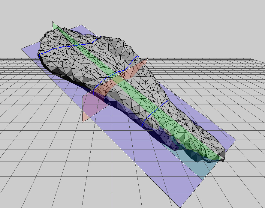
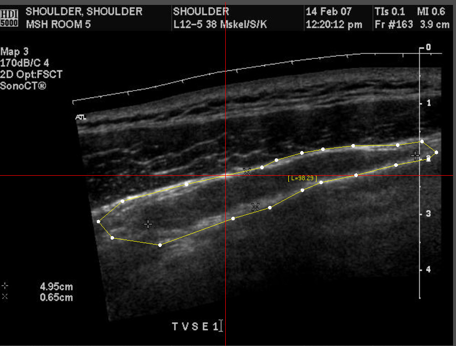
| Ultrasonographic assessment vs virtual (simulated) ultrasound | ||||||||
|
||||||||
| 3D Registration for muscle architecture (fitting cadaveric model to in-vivo ultrasonographic assessment) | |||||||||||
|
|||||||||||